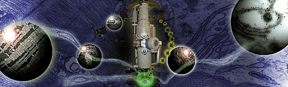
 This illustration by Adam Hartley in the Department of Cell Biology shows the power of the electron microscope, which can magnify cells and molecules a half a million times and opened a new universe to biologists in the 1930s. |
The power and possibilities of the electron microscope will be the focus of a program on Friday, Nov. 4, at the School of Medicine.
When Anton van Leeuwenhoek created his light microscope in the late 1600s, he ushered in a revolution in science, enabling humans to see a previously hidden world and to begin to understand the structure and function of microorganisms and individual cells magnified as many as 270 times.
Three centuries later, the invention of the electron microscope opened a new universe to scientists. By focusing a beam of fast-moving electrons -- rather than light -- on a sample, biologists could see cells and their parts half a million times larger, easily distinguishing the shapes of macromolecules for the first time.
It might seem that newer techniques such as magnetic resonance imaging, computed tomography and positron emission tomography would sideline older methods of visualizing life's smallest units -- that these newer methods, along with the sequencing of human and animal genomes, have given scientists enough new data about tissues, cells and their molecular components to render traditional and Electron Microscopy (EM) less relevant to today's biologists.
Not so, says Marc Pypaert, director of the EM facility at the School of Medicine and a research scientist in the Department of Cell Biology.
"It's really quite the opposite," says Pypaert. "There are now thousands of new proteins to be characterized in terms of function and localization. Combining the use of highly specific molecular probes, antibodies and gold conjugates with the high resolution of EM, scientists can now gain precise information on the location of proteins and other macromolecules within the cell that cannot be obtained using other techniques."
Pypaert and Ira S. Mellman, chair and Sterling Professor of Cell Biology and professor of immunobiology, will introduce the next Dean's Workshop at the School of Medicine, "Electron Microscopy in the Molecular Era: A World Inside the World You See," on Friday, Nov. 4, from 1:30 to 3:30 p.m. at the Anlyan Center auditorium, 300 Cedar St.
The workshop will feature talks by three medical school professors -- Graham Warren, Dr. Pietro De Camilli and Dr. Gerald Shulman, along with Dr. John Heuser, a visiting scientist from Washington University School of Medicine in St. Louis.
The speakers will describe their work with electron microscopy and describe what EM techniques can show scientists. Their respective topics are "From Golgi Vesicles to Golgi Stacks," "Endocytic Mechanisms at the Neuronal Synapse," "Mitochondrial Dysfunction and Type 2 Diabetes" and "Imaging Macromolecules in Their Cellular Context."
For his part, Pypaert was an early convert to EM, a technique invented in the early 1930s by two Germans, Max Knott and Ernst Ruska. As a postdoctoral fellow, Pypaert worked for several years in biochemistry and genetics labs and felt frustrated at "never seeing more than just a few bands on a gel or a sequence of letters on a piece of paper."
"Nothing beats the experience of first looking at a new protein under the electron microscope, or analyzing the phenotype of a new mutant under the beam of electrons," says Pypaert. "With a potential magnification factor of 500,000, even the smallest cell becomes the virtual size of a room, and the microscopist becomes like the cytonaut described by Nobel Prize winner Christian de Duve in his book 'A Guided Tour of the Living Cell,' exploring living matter from within."
The Dean's Workshop is free and open to the public and will be followed by a guided tour of the medical school's electron microscopy facility in Sterling Hall of Medicine. For details, visit www.med.yale.edu/workshops.
T H I S
Next Dean's Workshop focuses
on the electron microscope W E E K ' SS T O R I E S
![]() Yale expands its policy on sick leave
Yale expands its policy on sick leave![]()
![]() Researcher finds lower payments for treatment affect . . .
Researcher finds lower payments for treatment affect . . .![]()
![]() Faculty to study cell interactions in NIH project
Faculty to study cell interactions in NIH project![]()
![]() In trail guide, employee showcases her hometown's natural splendors
In trail guide, employee showcases her hometown's natural splendors![]()
![]() NIH grant supports network for research on preterm birth
NIH grant supports network for research on preterm birth![]()
![]() Library is a 'treasure-house of history,' says author
Library is a 'treasure-house of history,' says author![]()
![]() ENDOWED PROFESSORSHIPS
ENDOWED PROFESSORSHIPS
Anthropologist Burger is appointed as MacCurdy Professor![]()
Anthropologist Hill is designated as Stephenson Professor![]()
Political scientist Wallerstein is named as Saden Professor![]()
![]() University pays tribute to Robert Penn Warren . . .
University pays tribute to Robert Penn Warren . . .![]()
![]() Entertainer and activist to give Chubb Lecture
Entertainer and activist to give Chubb Lecture![]()
![]() Events mark century of Native American presence at Yale
Events mark century of Native American presence at Yale![]()
![]() Yale Law Journal launches an online companion publication
Yale Law Journal launches an online companion publication![]()
![]() Next Dean's Workshop focuses on the electron microscope
Next Dean's Workshop focuses on the electron microscope![]()
![]() Campus events celebrate German dramatist Friedrich Schiller
Campus events celebrate German dramatist Friedrich Schiller![]()
![]() Memorial service for Abraham S. Goldstein
Memorial service for Abraham S. Goldstein![]()
![]() A call for action
A call for action![]()
![]() Campus Notes
Campus Notes![]()
Bulletin Home|Visiting on Campus|Calendar of Events|In the News![]()
Bulletin Board|Classified Ads|Search Archives|Deadlines![]()
Bulletin Staff|Public Affairs|News Releases|
E-Mail Us|Yale Home